Dental X-rays are pictures of the teeth, bones, and soft tissues around them to help find problems with the teeth, mouth, and jaw. X-ray pictures can show cavities, hidden dental structures (such as wisdom teeth), and bone loss that cannot be seen during a visual examination. Dental X-rays may also be done as follow-up after dental treatments.
The following types of dental X-rays are commonly used.
The X-rays use small amounts of radiation.
- Bitewing X-rays show the upper and lower back teeth and how the teeth touch each other in a single view. These X-rays are used to check for decay between the teeth and to show how well the upper and lower teeth line up.
- Periapical X-rays show the entire tooth, from the exposed crown to the end of the root and the bones that support the tooth.
- Occlusal X-rays show the roof or floor of the mouth and are used to find extra teeth, teeth that have not yet broken through the gums, jaw fractures, a cleft in the roof of the mouth (cleft palate), cysts, abscesses, or growths. Occlusal X-rays may also be used to find a foreign object.
- Panoramic X-rays show a broad view of the jaws, teeth, sinuses, nasal area, and temporomandibular (jaw) joints. These X-rays do not find cavities. These X-rays do show problems such as impacted teeth, bone abnormalities, cysts, solid growths (tumors), infections, and fractures.
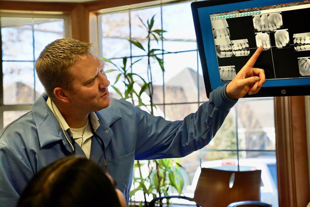
- Digital X-ray is a new method being used in some dental offices. A small sensor unit sends pictures to a computer to be recorded and saved.